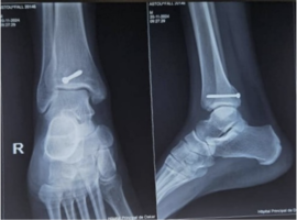
Background: Isolated posterior tibial margin fractures are exceptionally rare injuries, historically described among military parachutists. However, recent reports suggest a changing epidemiological profile, with an increasing number of cases occurring in civilian populations. This report describes a new civilian case and reviews the current literature to highlight recent epidemiological, diagnostic, and therapeutic developments.
Case Presentation: A 32-year-old woman sustained an isolated posterior tibial margin fracture following a fall while descending stairs. Standard radiographs suggested the diagnosis, which was confirmed by computed tomography (CT). CT demonstrated fragment displacement, comminution, and involvement of approximately 25% of the tibial articular surface. The patient underwent percutaneous reduction and screw fixation through a posteromedial approach. At two months, fracture union was achieved, with complete recovery of ankle motion and no residual pain.
Literature Review: A narrative review of the literature was conducted using PubMed/MEDLINE to identify publications on isolated posterior tibial margin fractures. Recent evidence indicates that although the injury mechanism remains unchanged, the epidemiological profile has evolved from predominantly military parachutists to civilian patients. CT has become the imaging modality of choice for confirming the diagnosis and guiding treatment decisions. Surgical fixation is generally recommended for displaced fractures involving a substantial portion of the articular surface.
Conclusion: Isolated posterior tibial margin fractures should no longer be regarded as injuries exclusive to military parachutists. Early recognition and systematic CT evaluation are essential to avoid missed diagnoses and to optimize treatment. Appropriate management based on CT findings is associated with excellent functional outcomes.
Keywords: Posterior tibial margin; Posterior malleolus; Isolated fracture; Computed tomography; Case report; Narrative review